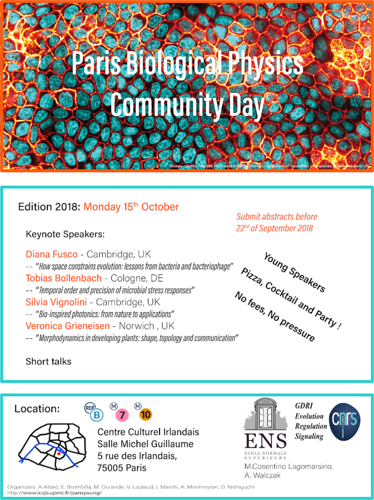
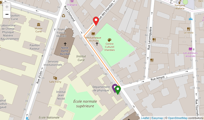
Monday 15th October 2018 Centre Culturel Irlandais @ 9am Salle Michel Guillaume - 5 rue des Irlandais, 75005 Paris
No fees. No pressure. All fun.
The Paris Biological Physics Community Day (PBPCD 2018) is a conference organized by young researchers of biological physics in the Paris area. We aim to bring together enthusiastic researchers in biophysics in the Paris area to create an opportunity for meeting and sharing knowledge.
The meeting is intended for researchers working in diverse areas of biophysics. It is going to be a day of conviviality and scientific enthusiasm, we envision to have a dynamic and informal atmosphere. In the program the talks of the invited speakers are interleaved with short presentations by young investigators.
The event is organized on behalf of GDRI (Groupement De Recherche International "Evolution, Regulation and Signaling") which also provides the funding.
No registration and no fees: lunch, coffee breaks and closing cocktail included!! Just come at the Centre Culturel Irlandais.
For any questions, contact us on social networks: Facebook Twitter
Keynote Speakers
Diana FuscoCambridge, UK
Tobias BollenbachKöln, Germany
Silvia VignoliniCambridge, UK
Veronica GrieneisenJohn Innes Centre, Norwich, UK
Program
8h30 - 9h15
Welcome coffee
9h15 - 10h45
Silvia Vignolini
Bio-inspired Photonics: from nature to applications
The most brilliant colours in nature are obtained by structuring transparent materials on the scale of the wavelength of visible light. By controlling/designing the dimensions of such nanostructures, it is possible to achieve extremely intense colourations over the entire visible spectrum without using pigments or colorants. Colour obtained through structure, namely structural colour, is widespread in the animal and plant kingdom. Such natural photonic nanostructures are generally synthesised in ambient conditions using a limited range of biopolymers. Given these limitations, an amazing range of optical structures exists: from very ordered photonic structures, to partially disordered, to completely random ones. In this seminar, I will introduce some striking example of natural photonic structures and review our recent advances to fabricate bio-mimetic photonic structures using the same material as nature. Biomimetic with cellulose-based architectures enables us to fabricate novel photonic structures using low cost materials in ambient conditions. Importantly, it also allows us to understand the biological processes at work during the growth of these structures in plants.
Manon Valet
Probing Molecular Transport in Printed Biomimetic Tissues
Cell-cell communication in biological tissues depends partly on the passive molecular transport through transmembrane channels. How does such diffusive transport lead to specific patterns in multicellular assemblies? We have recently addressed this question by studying the diffusive properties of fluorophores in biomimetic tissues. They consist of synthetic membranes known as Droplet Interface Bilayers (DIB) [1], connected by passive alpha-hemolysin pores. We have built linear arrays of DIBs using a recently developed droplet-on-demand technique [2]. We have measured the diffusion kinetics and its dependence with the pore concentration in the DIBs. Our results are fully captured by a model based on continuous time random walks and mean first passage times.
Nicolas Harmand
Merits and limits of surface and line tensions to understand the shape of epithelial cells
Building a physical framework to account for the shape of cells in epithelia is an important challenge to understand various biological processes, such as embryogenesis. As a PhD student, I aim to understand how surface tensions and line tensions shape epithelial cells using microstructured substrates, confocal fluorescence microscopy, force inference and theory. I culture epithelial cells on either flat or curved substrates and I explore the influence of the curvature of the substrate on the shape of these cells, especially the resulting thickness of the epithelial sheet. The model I propose to account for our measurements aims at computing the shape of individual cells within an epithelium using differentiated surface tensions for the different interfaces (cell-cell and cell-substrate surface tensions) and an apical line tension. I combine these measurements with force inference within the tissue using both the shape of cells in the epithelial sheet plane and the shape of the intercellular junctions within the thickness of the epithelium. I thus can infer which parameters determine the three dimensional shape of the cells and I can evaluate values for the different interfacial tensions by comparing the three-dimensional cell shape measurements with the prediction of the model and the prediction of force inference.
10h45 - 11h00
Flash Talks
Thomas Vourc'h
Surface diffusion of bacteria: from the particle to the biofilm
To survive under complex environments, living micro-organisms tend to self-organize in the form of a biofilm. This structure can be problematic -for instance when it protects pathogenic bacteria against drugs- and the basic mechanisms allowing for such a widespread organization are still debated. Here, we present a combined experimental and numerical study that describes the initial steps of biofilm formation in the case of the model cyanobacterium Synechocystis sp. PCC 6803. During the early stages of surface colonization, the observed intermittent motility, well-described by a continuous time random walk, slows down dramatically. As shown by the comparison of the diffusive dynamics of various mutants, this slowdown is due to the progressive coverage of the surface by the extracellular substances secreted by the bacteria. This growing two-dimensional network provides a specific structure, which constitutes a preliminary step for the biofilm formation. At long times, the morphology of the biofilm is provided by the microcolonies, composed by some bacteria bound to each other. The number of emerging microcolonies depends on the heterogeneous dynamics of the bacterial population. Finally, extensive numerical simulations suggest a complex interplay between growth, motility and attachment-detachment processes between cell, leading to different biofilm morphologies.
Alessandro Manacorda
Lattice model for fluctuating hydrodynamics in active matter
Active matter is every kind of physical system made of units capable of moving themselves through some self-propulsion mechanism: bacteria colonies, sheep herds, fish schools and bird flocks are among the most studied, and humans and robots as well can be considered active particles. Since the beginning of their observation, active systems have shown a rich phenomenology, especially in collective behaviors such as swarming, clustering, phase separation, giant number fluctuations and more. I present a model to derive the fluctuating hydrodynamics of non-overdamped self-propelled particles: inspired by granular lattice models and exclusion processes, we obtained the active hydrodynamic equations and reproduced the disordered, swarming and clustering state, as well as the amplitude of fluctuations. The transition between disorder and swarming in dilute systems can be predicted and confirmed by numerical simulations, shedding new light on the interplay between the control parameters and elucidating the relation between the active swarming phase and granular shear instability.
Marco Molari
Modeling affinity maturation: the role of antigen dosage
No medical procedure has been more successful in saving lives than vaccination. The core mechanism underlying vaccination is affinity maturation (AM), a darwinian evolution process whose outcome is a population of antibodies with high affinity for the administered pathogen. Recently, effort has been put into devising optimal immunization strategies. Examples include controlling the antigen (Ag) dosage and administration schedule or, in the case of mutable pathogens, by administering cocktails of different mutants. Following these approaches we introduce an analytical model for AM with which we aim to study the effect of Ag dosage on the outcome of immunization. Through the solution of an eigenvalue equation we find that our model presents different asymptotic phases according to the Ag concentration. In particular one phase at intermediate concentration appears to be optimal for developing high affinity antibodies. With a similar approach we characterize the acquisition of affinity towards mutants of the pathogen when immunizing against a wild-type, as a function of both Ag concentration and distance of mutant from the wild-type. Finally we simulate the variation of Ag concentration over time due to Ag consumption and Ag decay, and test different immunization schemes. In this setting as well we find an intermediate optimal Ag dosage.
Ido Lavi
Motility and waves in a hydrodynamic model of confined cell fragments
I will discuss a physical model of polarization, migration, and deformation by biological cell fragments confined between two parallel plates (Hele-Shaw cell). The fragment is described as a viscous droplet that contains a force-transducing solute. On the droplet's free boundary, this solute modulates an active traction force that offsets the Young-Laplace condition. It is shown that an increase in solute activity destabilizes a global translation-polarization mode, thereby prompting spontaneous symmetry breaking and motility. Higher activity begets Hopf bifurcations of morphological deformation modes - characterized by normal shape oscillations evolving in concert with alternating solute distributions of the Bessel type (both standing and traveling waves). Due to its simplicity, this model presents a paradigm of an over-damped hydrodynamic system that manifests slow inertial-like behaviors over diffusion timescales, akin to those exhibited by biological cells.
11h00 - 11h30
Coffee Break
11h30 - 13h00
Tobias Bollenbach
Temporal order and precision of microbial stress responses
Sudden stress often triggers diverse, temporally structured gene expression responses in microbes,but it is largely unknown how variable in time such responses are and if genes respond in the same temporal order in every single cell. We quantified timing variability of individual promoters responding to sublethal antibiotic stress using fluorescent reporters, microfluidics, and time-lapse microscopy. We identified lower and upper bounds that put definite constraints on timing variability, which varies strongly among promoters and conditions. Timing variability can be quantitatively rationalized with a model from queuing theory, which enables us to estimate the number of rate-limiting molecular steps underlying different responses. We found that just a few critical steps control some responses while others rely on dozens of steps. To probe connections between different stress responses, we then tracked the temporal order and response time correlations of promoter pairs in individual cells. Our results support that, when bacteria are exposed to the antibiotic nitrofurantoin, the ensuing oxidative stress and SOS responses are part of the same causal chain of molecular events. In contrast, under trimethoprim, the acid stress response and the SOS response are part of different chains of events running in parallel. Our approach reveals fundamental constraints on gene expression timing and provides new insights into the complexity of molecular events that underlie the timing of stress responses.
Maximilian Puelma Touzel
Inference of perturbed immune repertoire statistics enables precise-tracking of responding T-cell clones
The T-cell repertoire response to the yellow fever vaccine builds near total immunity. To quantify the response of this high dimensional stochastic system we developed a statistical model of differential T-cell proliferation and used it as a basis for inference from high-throughput receptor sequencing data obtained from individuals pre and post vaccination. We used replicate data to infer parameters of the experimental variation of clone sizes, and pre and post vaccination data to quantify the response and identify candidate clones responsive to the vaccine by their posterior expansion probability. While the learned replicate models suggest a near universal statistics for the experimental variability, the learned repertoire ensemble parameters vary temporally, consistent with the known timescales of yellow fever responses. Moreover, candidate clones identified by our method as contributing to this variation are experimentally validated, demonstrating its usefulness as a diagnostic tool in the clinic.
Rocio Espada
Infer repeat protein energetics from evolutionary information
Natural protein sequences contain a record of their history. A common constraint in a given protein family is the ability to fold to specific structures, and it has been shown possible to infer the main native ensemble by analyzing covariations in extant sequences. Still, many natural proteins that fold into the same structural topology show different stabilization energies, and these are often related to their physiological behavior. We proposed a quantitative model, derived from statistical physics, for the energetic variation given by sequence modifications in repeat proteins, systems for which the overall problem is simplified by their inherent symmetry. We explicitly accounted for single amino acid and pair-wise interactions and treated higher order correlations with a single term. In this talk I will show that the resulting evolutionary field captures higher order statistics of the protein family. The variations in the energetic scores of natural proteins is related to their experimental characterization and allows the prediction of the folding free energy change for several mutants. Finally, I will discuss the possibility of using this model to generate synthetic sequences that are statistically indistinguishable from the natural counterparts.
Morphodynamics in developing plants: shape, topology and communication
Just over a century ago, D'Arcy Thompson's published his canonical work "On Growth and Form", in which a new vision on how to address biological morphogenesis through quantitative principles was presented. It opened the door to mathematicians and physicists to start tackling problems of shape, form and dynamics in biology. I here wish to discuss the legacy of these ideas in a modern context of contemporary imaging and tracking techniques. I will focus on a cell shape misfit mentioned in his book, the plant pavement cell and the developing leaf. Through topological quantifications, we are able to infer how cellular decisions regarding divisions are made, and through high-throughput data imaging and analysis these findings can be verified. However, to unravel how ultimately intracellular polarity decisions lead to higher level patterning, we find that the field of morphometrics is still in need of tools and methods hat can better capture the developmental complexity. I will present a quantification technique we developed to fill this gap, coined LOCO-EFA. Together, our results lead to surprising new questions regarding patterning and decisions on cell polarity, division and growth.
Suzanne Ferte Fogel
Measuring an alternative cyclic electron flow within photosynthetic chain using fluorescence and spectroscopic tools
Most of the reduced carbon on Earth comes from photosynthesis. In this process, a crucial role is played by the transfer of electrons from H2O to CO2 through a chain of reactions called linear electron flow (LEF) and producing O2 and sugars. As early as the ‘50s, it has been proposed that an alternative mode, called cyclic electron flow (CEF), could take place concomitantly. This CEF seems essential for photosynthesis efficiency and operation but most of the initial questions are still largely debated. We know that several photosynthetic complexes are common to both LEF and CEF. But what are the proteins specifically involved in CEF pathway? What is its biological role and is it present in all phylogenetic clades? How is it regulated? This lack of consensus is mostly due to the complexity of CEF measurement: a cyclic flow does not provide any net product by definition. Several methods have been proposed but there is no general agreement within the community on a robust and precise method to evaluate CEF. In our work, we propose a simple approach to test (i) the existence of CEF and (ii) its relationship with LEF, in various microalgal species. We use chlorophyll fluorescence to measure the activity of a complex involved in LEF only and a time-resolved spectroscopic method (based on the Stark effect of photosynthetic pigments) to evaluate the activity of complexes involved in both LEF and CEF. Then we use a specific inhibitor of the LEF to progressively decrease LEF and we follow the evolution of LEF + CEF. By doing so, we can visualize the presence of CEF and understand the inter-relation between the two modes of electron transport. We could show that CEF does not occur in all phytoplankton species under non-stressful conditions and continuous illumination. When CEF is present, we observed very different relationship between CEF and LEF in different species, which we are now trying to relate to genetic or structural differences.
Laura Alaimo
Collective cell migration under a 3D spatial confinement
The growth of epithelial tissues in confined microenvironment is essential for the development of lumens in the human body. However most of the prior studies investigating the role of physical cues on collective cell migration employed two-dimensional (2D) flat culture systems that do not replicate out-of-place spatial confinements encountered in complex physiological environments. To address this issue, we studied the coordinated migration of epithelial cell sheets in microchannels of widths ranging from 100 to 300 µm for mimicking three-dimensional (3D) microenvironments. We observed that the cell density decreases from the rear to the front of the tissue, whereas the mean cell area conversely increases, independently of the channel dimension. Our findings show that the migration velocity increases with the channel widening but drops significantly with time. Interestingly, we demonstrate that the jamming transition from a solid-like to a fluid-like state is not controlled by the cell density but rather by the strengthening of cell adhesions. Altogether, our findings provide insights into the emerging migratory modes for epithelial migration and growth under 3D spatial confinement, which are reminiscent of the in vivo scenario.
16h00-16h30
Coffee Break
16h30-18h00
Diana Fusco
How space constrains evolution: lessons from bacteria and bacteriophage
Spatially growing populations are ubiquitous across scales, ranging from microbial biofilms in the soil to expanding tissues in developing organs and the spreading of diseases. In spatial settings, individuals experience inhomogeneities in the surrounding environment that result in different growth rates across the population. Since replication is necessary to transmit the genetic information from mother to daughter, the growth dynamics determined by the spatial constraints can deeply affect the spreading of mutations in a population and thus its evolution. Here, I will present two examples in which the simple spatial constraints associated with two-dimensional growth affect both the genetic diversity and the adaptation of microbial and viral populations, respectively. Population sequencing and fluorescence imaging show that microbial colonies exhibit an excess of mutational jackpot events compared to a well-mixed population of the same size. At the same time, they also carry a large number of rare mutations that are homogeneously distributed across the population and continuously generated during the growth process. A very different growth and evolutionary dynamics is instead observed in ecoliphage T7 two-dimensional plaques. Here the phage has to find an optimal incubation time, which provides a sufficient offspring number without excessively delaying its diffusion.
Salome Gutierrez-Ramos
Acoustic confinement of Escherichia coli: the impact on biofilm formation
Brownian or self-propelled particles in aqueous suspensions can be trapped by acoustic fields generated by piezoelectric transducers usually at frequencies in the megahertz. The obtained confinement allows the study of rich collective behaviours like clustering or spreading dynamics in microgravity-like conditions. The acoustic field induces the levitation of selfpropelled particles and provides secondary lateral forces to capture them at nodal planes. Here, we give a step forward in the field of confined active matter, reporting levitation experiments of bacterial suspensions of Escherichia coli. Clustering of living bacteria is monitored as a function of time, where different behaviours are clearly distinguished. Upon the removal of the acoustic signal, bacteria rapidly spread, impelled by their own swimming. Trapping of diverse bacteria phenotypes result in irreversible bacteria entanglements and in the formation of free-floating biofilms.
Daniele Conti
Propagating speed waves in flocks
An efficient collective response to external perturbations is one of the most striking abilities of a biological system. One of the crucial aspect of this phenomenon is given by the information transfer, and resulting propagation of signals, within the group. In this respect it is well-known the existence of density waves that propagate linearly on birds flock. However, most aspects of this phenomenon are still not fully captured by theoretical models. We present a new model for the propagation of speed fluctuations inside a flock, which is able to reproduce the observed density waves.